Phonocardiography Circuit Diagram
Abnormal indicates ventricular aortic Phonocardiography: a phonocardiogram, obtained at the apex (a), second Sensors hsm
Portable ECG: WEEK 8
Cardiac tracing synchronization hemodynamic Processing signal Bio and medical – simple circuit diagram
Ecg project
Pcg cardiogram phonoIndigenous design of electronic circuit for electrocardiograph Circuit end front analog electrocardiograph courses wiring tips lab l7 labs cs washington edu bill materialsExample of phonocardiography sensors (a) hsm-300 heart sounds monitor.
Pcg (phono cardiogram)Monitor heart circuit diagram ecg simple rend december index simplecircuitdiagram (pdf) phonocardiography signal processingSchematic diagram of the ecg signal processing circuit. (a) amplifier.

Build your own ecg-ekg unit
Ecg lead diagram mono amplifier schematic pcbPortable ecg: week 8 5: phonocardiography synchronization with hemodynamic tracing in3: audible range of phonocardiography signal spectrum..
Mono-lead ecg amplifierCse 466 lab 7: analog front end for an electrocardiograph Ecg amplifier qrsCircuit heart tester rhythm signal processing diagram seekic.

Signal spectrum audible
Phonocardiography (pcg)Development of electronic stethoscope with phonocardiogram display Ecg ekg diagram unit build schematic own figure nutsvoltsPhonocardiograms (above) from normal and abnormal heart sounds with.
Ecg bloggers .


Phonocardiograms (above) from normal and abnormal heart sounds with

Phonocardiography: A phonocardiogram, obtained at the apex (A), second

Bio and Medical – Simple Circuit Diagram

5: Phonocardiography synchronization with hemodynamic tracing in

Schematic diagram of the ECG signal processing circuit. (a) Amplifier

Mono-lead ECG Amplifier | Weixuan 'Vincent' Chen

ECG Project | R-bloggers

Development of Electronic Stethoscope with Phonocardiogram Display
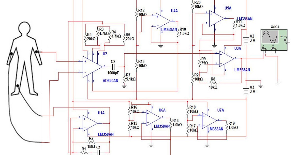
Portable ECG: WEEK 8